IX biO SS1 CHAP-TISSUE NOTES
Tissue: A groups of cells which is meant for a specific task is called tissue.
Tissue and Division of Labour: In complex organisms, different tasks are carried out by different organs and organ systems. Tissues are the first step towards division of labour in complex organisms.
Plant Tissues:Plant tissues are of two main types, viz. meristematic tissue and permanent tissue.
1 - Meristematic Tissue:-
Plant tissues in which cells keep on dividing are called meristematic tissue. Meristematic tissues are present in those parts of plants which keep on growing. Meristematic tissues are classified on the basis of their location. They are of following types:
a. Apical Meristem:- Apical meristem is present on root apex, stem apex, leaf buds and flower buds. They are responsible for growth in length, i.e. primary growth.
b. Lateral Meristem: Lateral meristem is present along the side of the stem. They are responsible for growth in girth, i.e. secondary growth.
c. Intercalary Meristem: Intercalary meristem is present at the base of leaf or internodes. They are present on either side of the node.
Since cells of meristematic tissue are highly active so they have dense cytoplasm. Vacuole is absent in these cells.
2 - Permanent Tissue:
Once the cells of meristematic tissue divide to a certain extent, they become specialized for a particular function. This process is called differentiation. Once differentiation is accomplished, the cells lose their capability to divide and the tissue becomes permanent tissue. Permanent tissues are of two types, simple permanent tissue and complex permanent tissue.
a) Simple Permanent Tissue: Simple permanent tissue is composed of similar types of cells. Simple permanent tissues are of three types, viz. parenchyma, collenchyma and sclerenchyma.
(1) Parenchyma: The cells of parenchyma have thin cell wall. They are loosely packed; with lot of intercellular spaces between them. Parenchyma makes the largest portion of a plant body. Parenchyma mainly works are packing material in plant parts. The main function of parenchyma is to provide support and to store food. In some plant parts, parenchyma has chlorophyll as well. In that case, parenchyma carries out photosynthesis and is then termed as chlorenchyma. In aquatic plants, large air cavities are present in parenchyma. This provides buoyancy to the plant, and
then the parenchyma is known as aerenchyma.

(2) Collenchyma: The cell wall of collenchyma is thickened at corners. Intercellular spaces are very few. Collenchymas provides some degree of structural rigidity with flexibility. Collenchymas is present in leaf stalk; below the epidermis. Due to this, the leaf talk can easily bend but does not break

(3) Sclerenchyma: The cell wall in sclerenchyma is highly thickened all around. The cells are dead and intercellular space is absent. Sclerenchyma provides structural rigidity to plant parts. Bark is composed of sclerenchyma. Another example of sclerenchyma can be seen in the coconut husk.
Stomata: The epidermis of leaves has small pores called stomata. A stoma is a composed of two guard cells which regulate the opening and closing of stoma. Stomata facilitates exchange of gases and transpiration.
(b) Complex Permanent Tissue:
Complex permanent tissue is composed of different types of cells. Complex permanent tissues are of two types, viz. xylem and phloem. Xylem and phloem together make the vascular bundle in plants.
1. Xylem: Xylem is composed of trachieds, vessels, xylem parenchyma and xylem fibres. The cells of xylem are dead; except the cells of xylem parenchyma. Trachieds and vessels are tubular structures and thus they provide a channel for conduction of water and minerals. Xylem fibre provides structural support to the tissue. Xylem parenchyma stores food.
2. Phloem: Phloem is composed of sieve tubes, companion cells, phloem fibre and phloem parenchyma. Sieve tubes are tubular cells with perforated walls. Sieve tubes are the conducting elements of phloem. Phloem is responsible for translocation of food in plants. The transport of food in phloem is a two way movement.
XYLEM & PHOLEM DIGRAM

Tissue----Animal Tissues
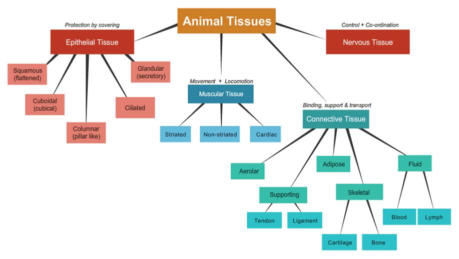
Animal tissues are of four types, viz. epithelial tissue, connective tissue, muscular tissue and nervous tissue.
Epithelial Tissue:
The epithelial tissue forms the covering or lining of most of the organs. The cells of epithelial tissue are tightly packed and form a continuous sheet. There is small amount of cementing materials between the cells and no intercellular space is present. Permeability of the epithelial tissue plays a great role in exchange of materials among various organs it also plays an important role in osmoregulation. All epithelial tissues are separated by the underlying tissue by an extracellular fibrous basement membrane. Epithelial tissues are of following types:
ANIMAL TISSUE DIGRAM
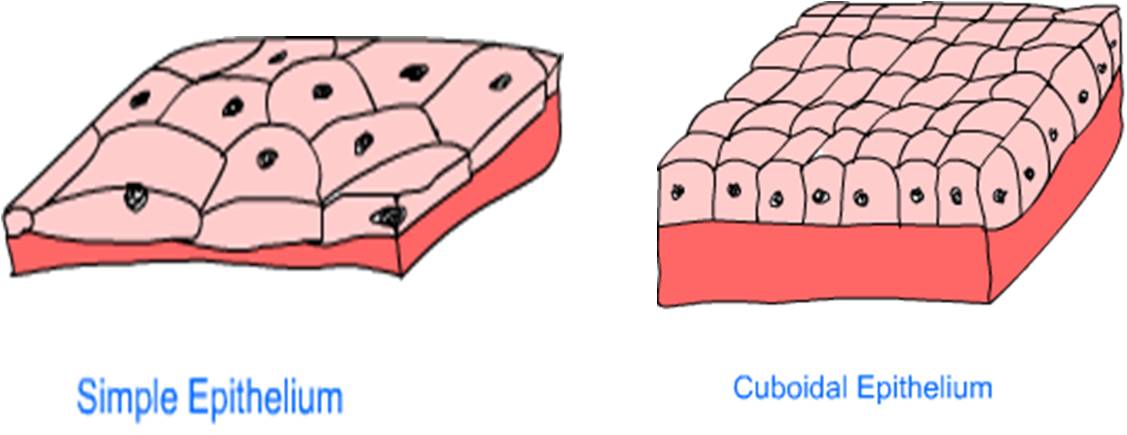
a. Simple Epithelium: The simple epithelium is composed of a single layer of cells. This type of epithelial tissue forms the lining of blood vessels and alveoli. Thin layer of cells facilitates exchange of substances; in such cases.
b. Cuboidal Epithelium:- The cells are cube-shaped in cuboidal epithelium. Linings of kidney tubules and ducts of salivary glands are composed of cuboidal epithelium. Cuboidal cells provide mechanical support. Cells of epithelium may play the role of secretion and then they are called glandular epithelium.
c. Columnar Epithelium:- Cells are column-shaped in columnar epithelium. Columnar epithelium facilitates secretion and absorption. For example; the lining of intestine is composed of columnar epithelium. In some organs, columnar epithelium has cilia present on the outer surface. Cilia facilitate movements of certain substances. The ciliated epithelium in the respiratory tract pushes the mucus forward.
d. Stratified Epithelium:- Cells of the stratified epithelium are in many layers. Skin is an example of stratified epithelium. Stratification of layers prevents wear and tear.
sIMPLE & COMPLEX EPITHELIUM
Connective Tissue:
The cells of a connective tissue are loosely scattered in a matrix. The matrix can be a fluid, jelly like, dense or rigid. The nature of matrix depends on the function a connective tissue serves. Following are the various connective tissues:
a. Areolar Connective Tissue: Areolar tissue is found between skin and muscles, around blood vessels and nerves and in bone marrow. Areolar tissue fills the gap between tissues and provides support. It also helps in repair of tissues.
b. Adipose Tissue: Adipose tissue is composed of fat globules. This tissue is found below the skin and beneath the organs. Adipose tissue provides insulation and works as a cushion.
c. Bone: Bone is mainly composed of osteoblasts. Bone makes the skeletal system. Skeletal system is responsible for providing structural framework to the body. It provides protection to important organs and facilitates movements.
d. Cartilage: Cartilage is mainly composed of chondrioblasts. Cartilage is present at the ends of articulatory bones. Cartilage is also present in external ear, bronchii, etc.
e. Blood: Blood is composed of blood cells, platelets and plasma. Blood plays an important role in transportation of various substances in the body. It also helps in osmoregulation and temperature control.
Muscular Tissue:
Muscular tissue is composed of muscle cells. Muscle cells are specialized cells which have the capability to contract and expand. Due to contraction and expansion, muscles facilitate various kinds of movements in the body. Muscular tissues are of three types:
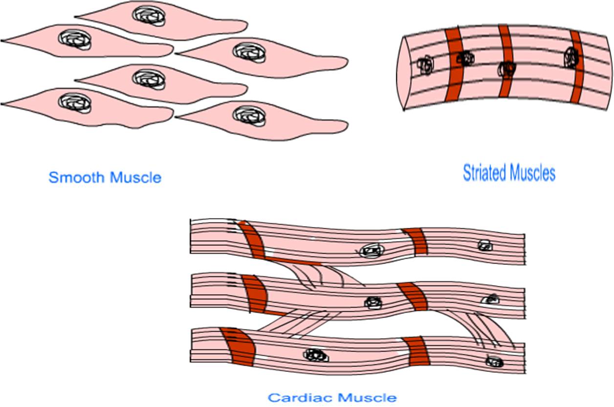
- Striated Muscles: The cells of striated muscles are in the form of long, unbranched fibres. Cells are multinucleate. Light and dark bands (striations) are present on muscle fibres; which gives the name striated muscles. Striated muscles are found in those organs where voluntary movement is possible, e.g. hands, legs, back, neck, etc.
- Smooth Muscles: The cells of smooth muscles are spindle shaped and each has one nucleus. Smooth muscle is found in those organs where involuntary movement is possible, e.g. alimentary canal.
- Cardiac Muscles: The cells of cardiac muscles are in the form of branched fibres. Striations are present and cells are uninucleate. These are found in the heart. Cardiac muscles are capable continuous contraction and relaxation throughout the life.
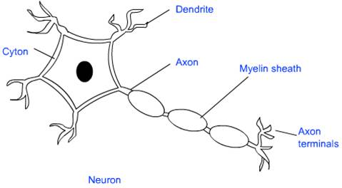
Nervous Tissue:
Nervous tissue makes the nervous system and is composed of specialized cells called neuron. A neuron can be divided into two distinct parts, viz. head and tail. The head is somewhat star-shaped and contains nucleus and some other cell organelles. This is called cyton. There are numerous hair-like outgrowths coming out of the cyton. These are called dendrites. The tail ends in axon terminals. Dendrites receive the nerve impulse, while axon relays the nerve signals
Nervous Tissues
1. These are composed of nerve cells which are called neurons.
2. Each neuron consists of a cell body, axon and dendrites.
3. These productive nerve impulse to conduct messages.
4. By this tissue, different body parts have coordination with each other.
5. This tissue also forms brain and spinal cord